Saturday, April 3, 2010
X-ray machine
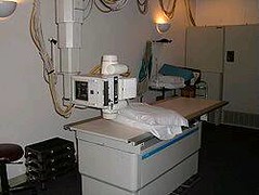
X-ray machine An X-ray generator is a device used to generate X-rays. These devices are commonly used by radiographers to acquire an x- ray image of the inside of an object (as in medicine or non- destructive testing) but they are also used in sterilization or fluorescence.An X-ray imaging system consists of a X-ray source or generator ( X-ray tube), an image detection system which can be either a film (analog technology) or a digital capture system, and a PACS. X-ray photons are produced by an electron beam that is accelerated to a very high speed and strikes a target. The electrons that make up the beam are emitted from a heated cathode filament. The electrons are then focused and accelerated by an electrical field towards an angled anode target. The point where the electron beam strikes the target is called the focal spot. Most of the kinetic energy contained in the electron beam is converted to heat, but around 1% of the energy is converted into X-ray photons, the excess heat is dissipated via a heat sink. At the focal spot, X-ray photons are emitted in all directions from the target surface, the highest intensity being around 60deg to 90deg from the beam due to the angle of the anode target to the approaching X-ray photons. There is a small round window in the X-ray tube directly above the angled target. This window allows the X-ray to exit the tube with little attenuation while maintaining a vacuum seal required for the X-ray tube operation. X-ray machines work by applying controlled voltage and current to the X-ray tube, which results in a beam of X-rays. The beam is projected on matter. Some of the X-ray beam will pass through the object, while some are absorbed. The resulting pattern of the radiation is then ultimately detected by a detection medium including rare earth screens (which surround photographic film), semiconductor detectors, or X-ray image intensifiers. Detection In healthcare applications in particular, the x-ray detection system rarely consists of the detection medium. For example, a typical stationary radiographic x- ray machine also includes an ion chamber and grid. The ion chamber is basically a hollow plate located between the detection medium and the object being imaged. It determines the level of exposure by measuring the amount of x-rays that have passed through the electrically charged, gas-filled gap inside the plate. This allows for minimization of patient radiation exposure by both ensuring that an image is not underdeveloped to the point the exam needs to be repeated and ensuring that more radiation than needed is not applied. The grid is usually located between the ion chamber and object and consists of many aluminum slats stacked next to each other (resembling a polaroid lens). In this manner, the grid allows straight x-rays to pass through to the detection medium but absorbs reflected x-rays. This improves image quality by preventing scattered (non- diagnostic) x-rays from reaching the detection medium, but using a grid creates higher exam radiation doses overall. Images taken with such devices are known as X-ray photographs or radiographs. X-ray machines are used in health care for visualising bone structures and other dense tissues such as tumours. Non- medicial applications include security and material analysis.The two main fields in which x- ray machines are used in medicine are radiography and dentistry. Radiography is used for fast, highly penetrating images, and is usually used in areas with a high bone content. Some forms of radiography include: orthopantomogram — a panoramic x-ray of the jaw showing all the teeth at once mammography — x-rays of breast tissue tomography — x-ray imaging in sections Radiotherapy — the use of x-ray radiation to treat malignant cancer cells, a non-imaging application Fluoroscopy is used in cases where real-time visualization is necessary (and is most commonly encountered in everyday life at airport security). Some medical applications of fluorography include: angiography — used to examine blood vessels in real time barium enema — a procedure used to examine problems of the colon and lower gastrointestinal tract barium swallow — similar to a barium enema, but used to examine the upper gastroinstestional tract biopsy — the removal of tissue for examination X-rays are highly penetrating, ionizing radiation, therefore X- ray machines are used to take pictures of dense tissues such as bones and teeth. This is because bones absorb the radiation more than the less dense soft tissue. X-rays from a source pass through the body and onto a photographic cassette. Areas where radiation is absorbed show up as lighter shades of grey (closer to white). This can be used to diagnose broken or fractured bones. In fluoroscopy, imaging of the digestive tract is done with the help of a radiocontrast agent such as barium sulfate, which is opaque to X-rays.
Subscribe to:
Post Comments (Atom)
No comments:
Post a Comment