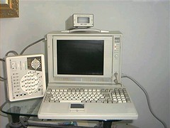
Electroencephalography machine Electroencephalography (EEG) is the recording of electrical activity along the scalp produced by the firing of neurons within the brain.In clinical contexts, EEG refers to the recording of the brain's spontaneous electrical activity over a short period of time, usually 20 –40 minutes, as recorded from multiple electrodes placed on the scalp. In neurology, the main diagnostic application of EEG is in the case of epilepsy, as epileptic activity can create clear abnormalities on a standard EEG study.A secondary clinical use of EEG is in the diagnosis of coma, encephalopathies, and brain death. EEG used to be a first-line method for the diagnosis of tumors, stroke and other focal brain disorders, but this use has decreased with the advent of anatomical imaging techniques such as MRI and CT. Derivatives of the EEG technique include evoked potentials (EP), which involves averaging the EEG activity time-locked to the presentation of a stimulus of some sort (visual, somatosensory, or auditory). Event-related potentials refer to averaged EEG responses that are time-locked to more complex processing of stimuli; this technique is used in cognitive science, cognitive psychology, and psychophysiological research.The electrical activity of the brain can be described in spatial scales from the currents within a single dendritic spine to the relatively gross potentials that the EEG records from the scalp, much the same way that economics can be studied from the level of a single individual's personal finances to the macro- economics of nations. Neurons, or nerve cells, are electrically active cells which are primarily responsible for carrying out the brain's functions. Neurons create action potentials, which are discrete electrical signals that travel down axons and cause the release of chemical neurotransmitters at the synapse, which is an area of near contact between two neurons. This neurotransmitter then activates a receptor in the dendrite or body of the neuron that is on the other side of the synapse, the post-synaptic neuron. The neurotransmitter, when combined with the receptor, typically causes an electrical current within the dendrite or body of the post- synaptic neuron. Thousands of post-synaptic currents from a single neuron's dendrites and body then sum up to cause the neuron to generate an action potential. This neuron then synapses on other neurons, and so on. EEG reflects correlated synaptic activity caused by post-synaptic potentials of cortical neurons. The ionic currents involved in the generation of fast action potentials may not contribute greatly to the averaged field potentials representing the EEG . More specifically, the scalp electrical potentials that produce EEG are generally thought to be caused by the extracellular ionic currents caused by dendritic electrical activity, whereas the fields producing magnetoencephalographic signals are associated with intracellular ionic currents.The electric potentials generated by single neurons are far too small to be picked by EEG or MEG.EEG activity therefore always reflects the summation of the synchronous activity of thousands or millions of neurons that have similar spatial orientation, radial to the scalp. Currents that are tangential to the scalp are not picked up by the EEG. The EEG therefore benefits from the parallel, radial arrangement of apical dendrites in the cortex. Because voltage fields fall off with the fourth power of the radius, activity from deep sources is more difficult to detect than currents near the skull. Scalp EEG activity shows oscillations at a variety of frequencies. Several of these oscillations have characteristic frequency ranges, spatial distributions and are associated with different states of brain functioning (e.g., waking and the various sleep stages). These oscillations represent synchronized activity over a network of neurons. The neuronal networks underlying some of these oscillations are understood (e.g., the thalamocortical resonance underlying sleep spindles), while many others are not (e.g., the system that generates the posterior basic rhythm). Research that measures both EEG and neuron spiking finds the relationship between the two is complex with the power of surface EEG only in two bands that of gamma and delta relating to neuron spike activity.A routine clinical EEG recording typically lasts 20 –30 minutes (plus preparation time) and usually involves recording from 25 scalp electrodes. Routine EEG is typically used in the following clinical circumstances: to distinguish epileptic seizures from other types of spells, such as psychogenic non- epileptic seizures , syncope (fainting), sub- cortical movement disorders and migraine variants. to differentiate "organic" encephalopathy or delirium from primary psychiatric syndromes such as catatonia to serve as an adjunct test of brain death to prognosticate, in certain instances, in patients with coma to determine whether to wean anti-epileptic medications At times, a routine EEG is not sufficient, particularly when it is necessary to record a patient while he/she is having a seizure. In this case, the patient may be admitted to the hospital for days or even weeks, while EEG is constantly being recorded (along with time-synchronized video and audio recording). A recording of an actual seizure (i.e., an ictal recording, rather than an inter- ictal recording of a possibly epileptic patient at some period between seizures) can give significantly better information about whether or not a spell is an epileptic seizure and the focus in the brain from which the seizure activity emanates. Epilepsy monitoring is typically done: to distinguish epileptic seizures from other types of spells, such as psychogenic non- epileptic seizures , syncope (fainting), sub- cortical movement disorders and migraine variants. to characterize seizures for the purposes of treatment to localize the region of brain from which a seizure originates for work-up of possible seizure surgery Additionally, EEG may be used to monitor certain procedures: to monitor the depth of anesthesia as an indirect indicator of cerebral perfusion in carotid endarterectomy to monitor amobarbital effect during the Wada test EEG can also be used in intensive care units for brain function monitoring: to monitor for non-convulsive seizures/non-convulsive status epilepticus to monitor the effect of sedative/anesthesia in patients in medically induced coma (for treatment of refractory seizures or increased intracranial pressure) to monitor for secondary brain damage in conditions such as subarachnoid hemorrhage (currently a research method) If a patient with epilepsy is being considered for resective surgery, it is often necessary to localize the focus (source) of the epileptic brain activity with a resolution greater than what is provided by scalp EEG. This is because the cerebrospinal fluid, skull and scalp smear the electrical potentials recorded by scalp EEG. In these cases, neurosurgeons typically implant strips and grids of electrodes (or penetrating depth electrodes) under the dura mater, through either a craniotomy or a burr hole. The recording of these signals is referred to as electrocorticography (ECoG), subdural EEG (sdEEG) or intracranial EEG (icEEG)--all terms for the same thing. The signal recorded from ECoG is on a different scale of activity than the brain activity recorded from scalp EEG. Low voltage, high frequency components that cannot be seen easily (or at all) in scalp EEG can be seen clearly in ECoG. Further, smaller electrodes (which cover a smaller parcel of brain surface) allow even lower voltage, faster components of brain activity to be seen. Some clinical sites record from penetrating microelectrodes.
No comments:
Post a Comment