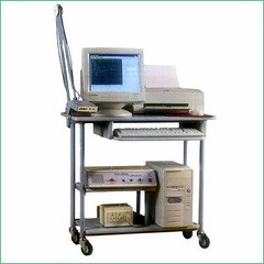
EMG machine Electromyography (EMG) is a technique for evaluating and recording the electrical activity produced by skeletal muscles.[1] EMG is performed using an instrument called an electromyograph, to produce a record called an electromyogram. An electromyograph detects the electrical potential generated by muscle cells[2] when these cells are electrically or neurologically activated. The signals can be analyzed to detect medical abnormalities, activation level, recruitment order or to analyze the biomechanics of human or animal movement. Electrical characteristics The electrical source is the muscle membrane potential of about -90 mV.[3] Measured EMG potentials range between less than 50 μV and up to 20 to 30 mV, depending on the muscle under observation. Typical repetition rate of muscle motor unit firing is about 7 –20 Hz, depending on the size of the muscle (eye muscles versus seat (gluteal) muscles), previous axonal damage and other factors. Damage to motor units can be expected at ranges between 450 and 780 mV. There are two kinds of EMG in widespread use: surface EMG and intramuscular (needle and fine- wire) EMG. To perform intramuscular EMG, a needle electrode or a needle containing two fine-wire electrodes is inserted through the skin into the muscle tissue. A trained professional (most often a physiatrist or neurologist) observes the electrical activity while inserting the electrode. The insertional activity provides valuable information about the state of the muscle and its innervating nerve. Normal muscles at rest make certain, normal electrical sounds when the needle is inserted into them. Then the electrical activity when the muscle is at rest is studied. Abnormal spontaneous activity might indicate some nerve and/or muscle damage. Then the patient is asked to contract the muscle smoothly. The shape, size, and frequency of the resulting motor unit potentials are judged. Then the electrode is retracted a few millimeters, and again the activity is analyzed until at least 10 –20 units have been collected. Each electrode track gives only a very local picture of the activity of the whole muscle. Because skeletal muscles differ in the inner structure, the electrode has to be placed at various locations to obtain an accurate study. Intramuscular EMG may be considered too invasive or unnecessary in some cases. Instead, a surface electrode may be used to monitor the general picture of muscle activation, as opposed to the activity of only a few fibres as observed using a intramuscular EMG. This technique is used in a number of settings; for example, in the physiotherapy clinic, muscle activation is monitored using surface EMG and patients have an auditory or visual stimulus to help them know when they are activating the muscle ( biofeedback). A motor unit is defined as one motor neuron and all of the muscle fibers it innervates. When a motor unit fires, the impulse (called an action potential) is carried down the motor neuron to the muscle. The area where the nerve contacts the muscle is called the neuromuscular junction, or the motor end plate. After the action potential is transmitted across the neuromuscular junction, an action potential is elicited in all of the innervated muscle fibers of that particular motor unit. The sum of all this electrical activity is known as a motor unit action potential (MUAP). This electrophysiologic activity from multiple motor units is the signal typically evaluated during an EMG. The composition of the motor unit, the number of muscle fibres per motor unit, the metabolic type of muscle fibres and many other factors affect the shape of the motor unit potentials in the myogram. Nerve conduction testing is also often done at the same time as an EMG to diagnose neurological diseases. Some patients can find the procedure somewhat painful, whereas others experience only a small amount of discomfort when the needle is inserted. The muscle or muscles being tested may be slightly sore for a day or two after the procedure.
No comments:
Post a Comment