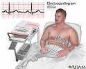
ECG machine & graph Electrocardiography (ECG or EKG) is a transthoracic interpretation of the electrical activity of the heart over time captured and externally recorded by skin electrodes. It is a noninvasive recording produced by an electrocardiographic device. The etymology of the word is derived from electro, because it is related to electrical activity, cardio, Greek for heart, and graph, a Greek root meaning "to write". Different types of ECGs can be referred to by the number of leads that are recorded, for example 3-lead, 5-lead or 12-lead ECGs (sometimes simply "a 12- lead"). A 12-lead ECG is one in which 12 different electrical signals are recorded at approximately the same time and will often be used as a one-off recording of an ECG, typically printed out as a paper copy. 3- and 5-lead ECGs tend to be monitored continuously and viewed only on the screen of an appropriate monitoring device, for example during an operation or whilst being transported in an ambulance. There may, or may not be any permanent record of a 3-lead ECG depending on the equipment used. The ECG works by detecting and amplifying the tiny electrical changes on the skin that are caused when the heart muscle "depolarises" during each heart beat. At rest, each muscle fibre has a charge across it's outer wall, or cell mambrane. Reducing this charge towards zero is called de-polarisation, which is what signals it to contract. Electrodes on different sides of the heart measure the activity of different parts of the heart muscle. An ECG displays the voltage between pairs of these electrodes. This display indicates the overall rhythm of the heart and weaknesses in different parts of the heart muscle. A healthy heart will have a characeristic pattern of electrical change, reflecting the orderly passage of depolarisation from the sinoatrial node, through the intrinsic conduction pathways and into the ventricles. It will also show when the negative charge resets (repolarisation). It is the best way to measure and diagnose abnormal rhythms of the heart, particularly abnormal rhythms caused by damage to the conductive tissue that carries electrical signals, or abnormal rhythms caused by electrolyte imbalances. In a myocardial infarction (MI), the ECG can identify if the heart muscle has been damaged in specific areas, though not all areas of the heart are covered. The ECG cannot reliably measure the pumping ability of the heart, for which ultrasound-based ( echocardiography) or nuclear medicine tests are used. It is possible to be dead with a normal ECG signal (a condition known as pulseless electrical activity).
No comments:
Post a Comment