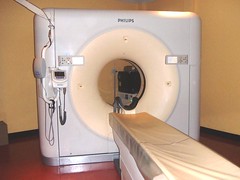
CT scanner Computed tomography (CT) is a medical imaging method employing tomography created by computer processing. Digital geometry processing is used to generate a three-dimensional image of the inside of an object from a large series of two- dimensional X-ray images taken around a single axis of rotation. CT produces a volume of data which can be manipulated, through a process known as "windowing", in order to demonstrate various bodily structures based on their ability to block the X-ray/Röntgen beam. Although historically the images generated were in the axial or transverse plane, orthogonal to the long axis of the body, modern scanners allow this volume of data to be reformatted in various planes or even as volumetric (3D) representations of structures. Although most common in medicine, CT is also used in other fields, such as nondestructive materials testing. Another example is the DigiMorph project at the University of Texas at Austin which uses a CT scanner to study biological and paleontological specimens. Usage of CT has increased dramatically over the last two decades. An estimated 72 million scans were performed in the United States in 2007. Terminology The word "tomography" is derived from the Greek tomos (slice) and graphein (to write). Computed tomography was originally known as the "EMI scan" as it was developed at a research branch of EMI, a company best known today for its music and recording business. It was later known as computed axial tomography (CAT or CT scan) and body section röntgenography. Although the term "computed tomography" could be used to describe positron emission tomography and single photon emission computed tomography , in practice it usually refers to the computation of tomography from X-ray images, especially in older medical literature and smaller medical facilities. In MeSH, "computed axial tomography" was used from 1977 –79, but the current indexing explicitly includes "X-ray" in the title.Since its introduction in the 1970s, CT has become an important tool in medical imaging to supplement X-rays and medical ultrasonography. It has more recently begun to also be used for preventive medicine or screening for disease, for example CT colonography for patients with a high risk of colon cancer. A number of institutions offer full-body scans for the general population. However, this is a controversial practice, given its lack of proven benefit, cost, radiation exposure, and the risk of finding 'incidental' abnormalities that may trigger additional investigations.
No comments:
Post a Comment